The knee is one of the most vital and complex joints in the human body, allowing us to perform a wide range of movements, including walking, running, jumping, and squatting. It serves as a crucial connection between the thigh bone (femur) and the shin bone (tibia), providing stability, flexibility, and weight-bearing support. Understanding the anatomy of the knee is essential for comprehending its function, common injuries, and the importance of maintaining knee health.
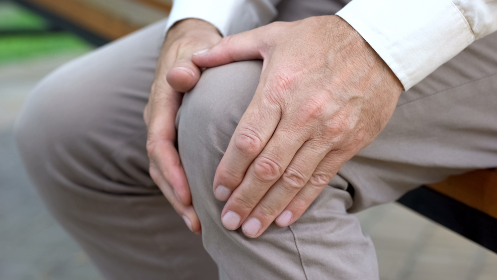
Due to its complexity, knee injuries can occur which can cause knee pain, weakness in the knee or even arthritis. Ongoing knee problems can create difficulties including walking, standing up and other movements involving the knee.
In this article, we will delve into the intricate structure of the knee joint and explore its various components.
Bones of the Knee
The knee joint is formed by the articulation of three bones: the femur, tibia, and patella (kneecap). Let's take a closer look at each of these bones:
- Femur (Thigh Bone): The femur is the longest and strongest bone in the body. Its rounded lower end, known as the femoral condyles, forms the upper part of the knee joint.
- Tibia (Shin Bone): The tibia is the larger of the two lower leg bones. It connects with the femur to form the lower part of the knee joint. The top surface of the tibia, called the tibial plateau, provides stability to the joint.
- Patella (Kneecap): The patella is a small, triangular bone located in front of the knee joint. It acts as a protective shield for the joint and improves the mechanical advantage of the thigh muscles by increasing leverage during movements like walking and running.
Ligaments of the Knee
Ligaments are strong bands of connective tissue that hold the bones together and provide stability to the knee joint. There are four main ligaments in the knee:
- Anterior Cruciate Ligament (ACL): The ACL runs diagonally inside the knee, preventing the tibia from sliding forward and providing rotational stability.
- Posterior Cruciate Ligament (PCL): The PCL is located at the back of the knee joint and prevents the tibia from sliding backward.
- Medial Collateral Ligament (MCL): The MCL is found on the inner side of the knee, providing stability against forces that push the knee inward.
- Lateral Collateral Ligament (LCL): The LCL is located on the outer side of the knee, providing stability against forces that push the knee outward.
Cartilage of the Knee
Cartilage plays a crucial role in the knee joint by cushioning the bones and allowing for smooth and pain-free movement. There are two types of cartilage in the knee:
- Articular Cartilage: Articular cartilage covers the ends of the femur, tibia, and the back of the patella. It provides a smooth and low-friction surface for the bones to glide over during movement.
- Meniscus: The menisci are two C-shaped pieces of cartilage located between the femur and tibia. They act as shock absorbers, distribute weight, and help stabilize the knee. The medial meniscus is on the inner side of the knee, while the lateral meniscus is on the outer side.
Muscles and Tendons of the KneeI
Several muscles and tendons surround the knee joint, enabling movement and providing strength and stability. Let's explore some key muscles and tendons:
- Quadriceps: The quadriceps are a group of muscles located in the front of the thigh. They include the rectus femoris, vastus lateralis, vastus medialis, and vastus intermedius. The quadriceps muscles straighten the knee and are essential for activities like walking, running, and jumping.
- Hamstrings: The hamstrings are a group of muscles located at the back of the thigh. They include the biceps femoris, semitendinosus, and semimembranosus. The hamstrings help flex the knee and are involved in activities like bending the knee, walking, and running.
- Patellar Tendon: The patellar tendon, also known as the kneecap tendon, connects the patella to the tibia. It plays a vital role in the extension of the knee joint, allowing for movements like jumping and kicking.
- Quadriceps Tendon: The quadriceps tendon connects the quadriceps muscles to the patella. It assists in the extension of the knee and helps stabilize the patella during movement.
Synovium and Synovial Fluid
The knee joint is surrounded by a synovial membrane, which produces synovial fluid. The synovium helps reduce friction within the joint and nourishes the cartilage. The synovial fluid acts as a lubricant, allowing smooth movement of the joint and reducing wear and tear on the cartilage.
Common Knee Injuries
The knee is prone to various injuries due to its complex structure and the significant forces it experiences during physical activities. Common knee injuries include:
- Anterior Cruciate Ligament (ACL) Tears: ACL tears often occur during sports activities and can cause instability and swelling in the knee.
- Meniscus Tears: Meniscus tears can result from sudden twisting or impact to the knee and can cause pain, swelling, and limited mobility.
- Patellar Tendonitis: Patellar tendonitis, also known as jumper's knee, is an overuse injury that causes pain and inflammation in the patellar tendon.
- Knee Osteoarthritis: Knee osteoarthritis is a degenerative condition that causes the cartilage in the knee joint to wear down over time, leading to pain, stiffness, and reduced mobility.
Conclusion
Our knees are truly remarkable structures that allow us to lead an active lifestyle and experience the world around us. However, knee injuries can be debilitating and hinder our ability to move freely with ease. Taking proper care of our knees is a key element in living a healthy, balanced life. Understanding the anatomy of the knee joint, using precautionary measures during physical activities, and consulting a healthcare professional if necessary are all components of keeping our knees healthy in the long run. Now that you have learned more about the intricate inner workings of your knee joint, take this newfound knowledge and put it to good use!
If you have been struggling with knee pain, it may be time for an evaluation. Dr. Brent Ungar treats knee pain and injuries using a variety of non-surgical techniques. Contact Belden Village of Chiropractic & Wellness Center to set up an appointment.
References:
https://orthoinfo.aaos.org/en/diseases--conditions/knee-anatomy.
https://www.physio-pedia.com/Knee_Joint_Anatomy